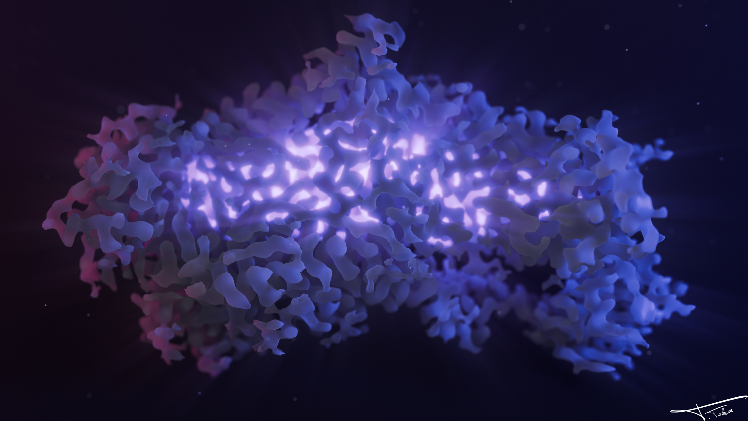
Illustration of a cryo-EM map
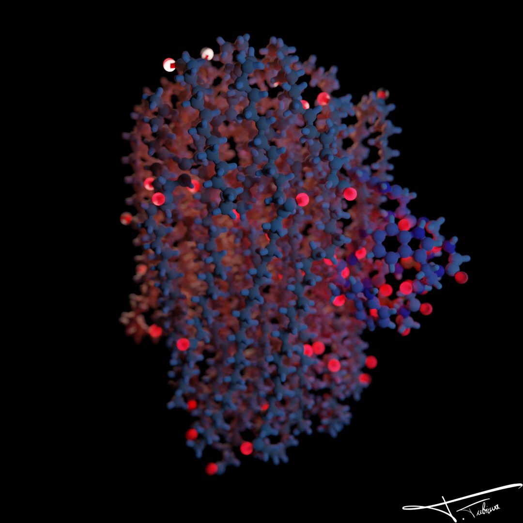
Illustration of a cryo-EM mapA cryo-EM density map, illustrated with some lighting effects.
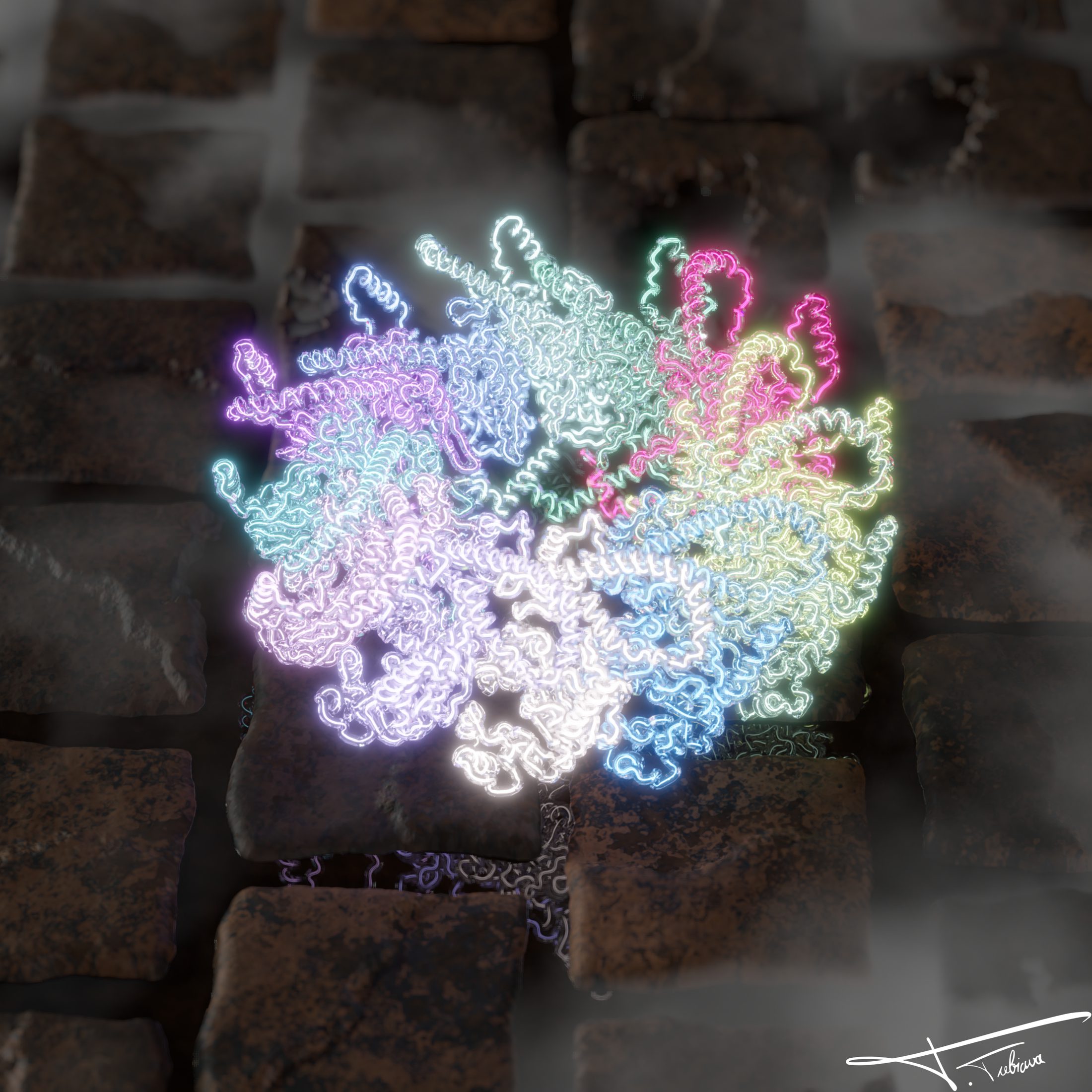
MetY dodecamer
MetY dodecamerIllustration of the HEV ORF1 MetY dodecamer with atiled floor.
ORF1 AlphaFold model animation, Goodsell styleanimation of our ORF1 Alphafold model, colored per PLDDT in the style of David Goodsell.
Norovirus in a bottle β€” PDB 4V60
Norovirus in a bottle β€” PDB 4V60The VP1 dodecamer, preserved like a museum piece.
I2BC, molecule by moleculeThe institute's name emerges from a sea of molecules.
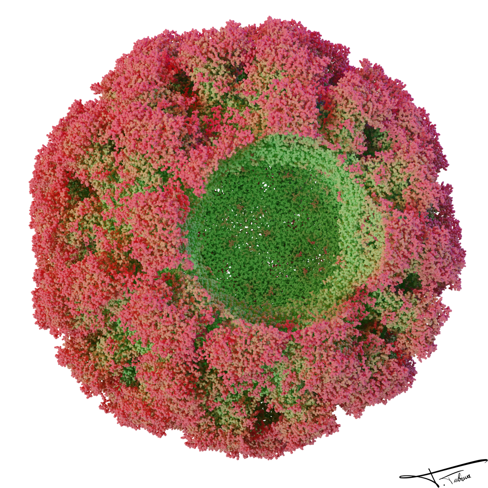
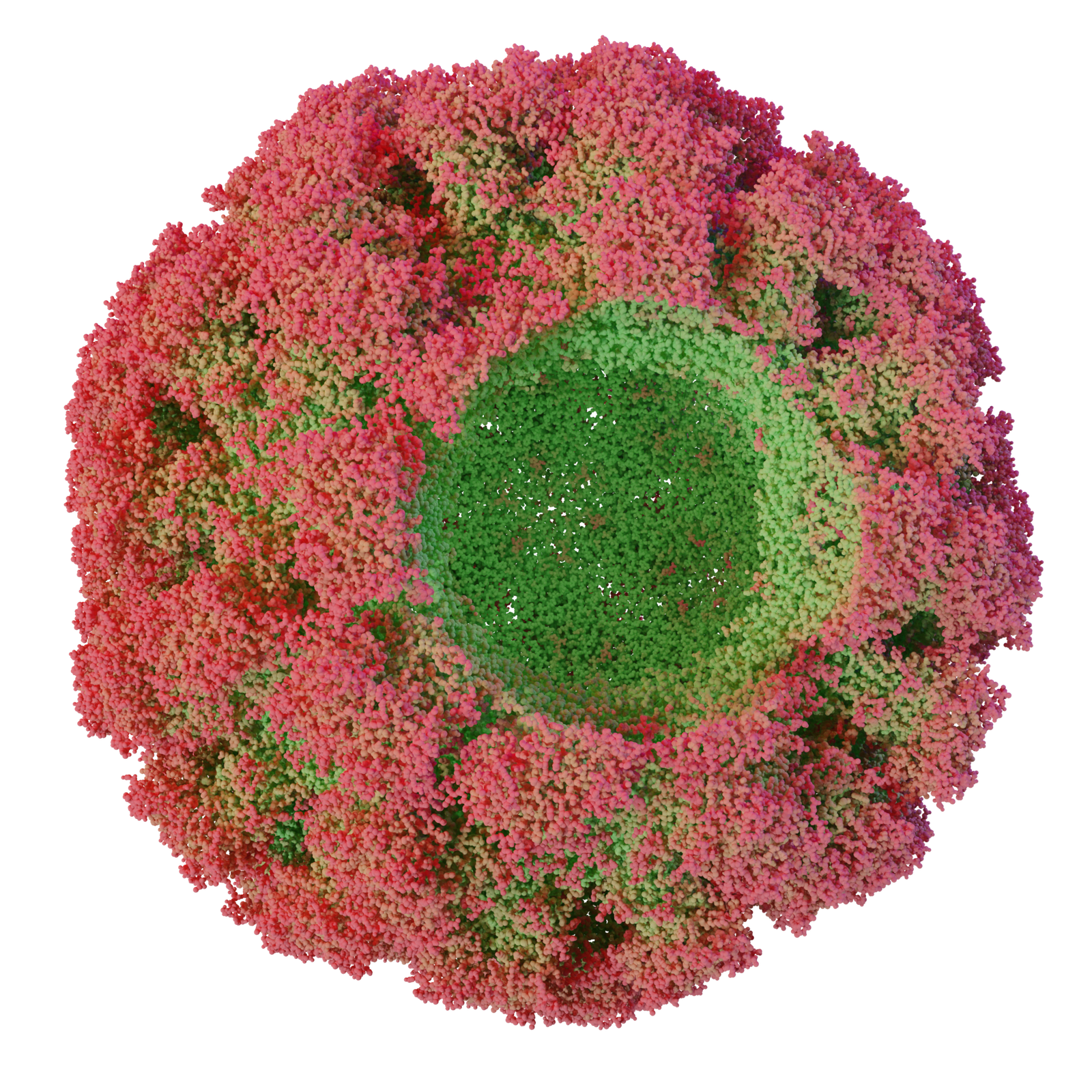
Viral capsid
Viral capsidAtomic surface with an open portal, colored by depth.
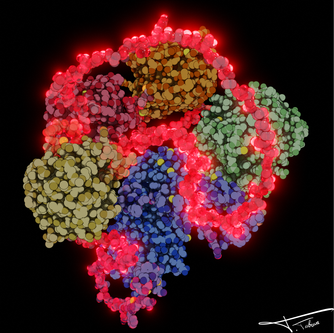
Neon dodecamer β€” lights onSubunits ignite one after another.
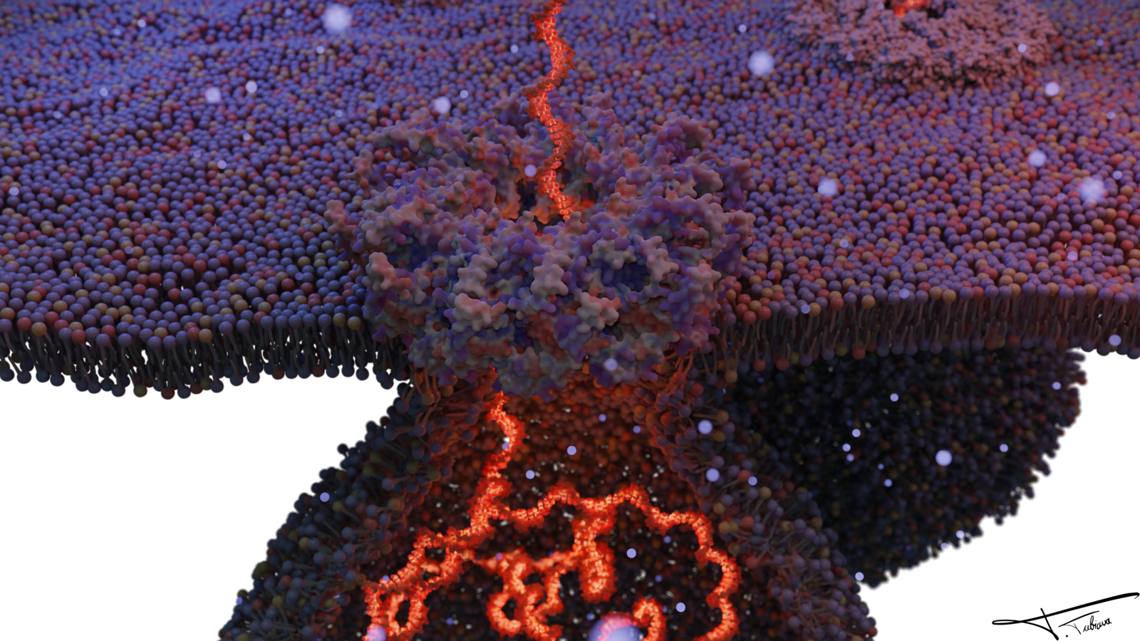
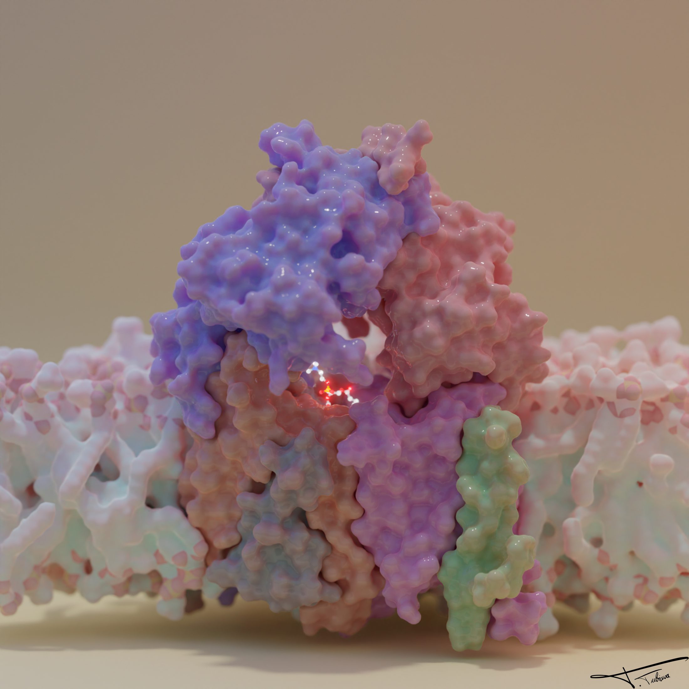
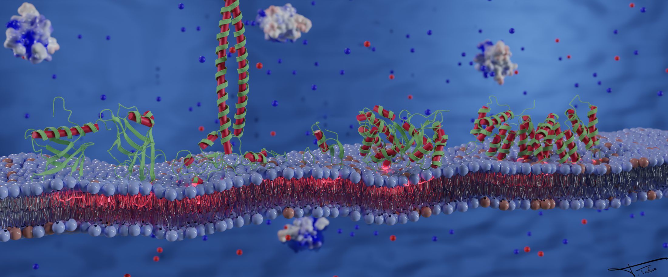
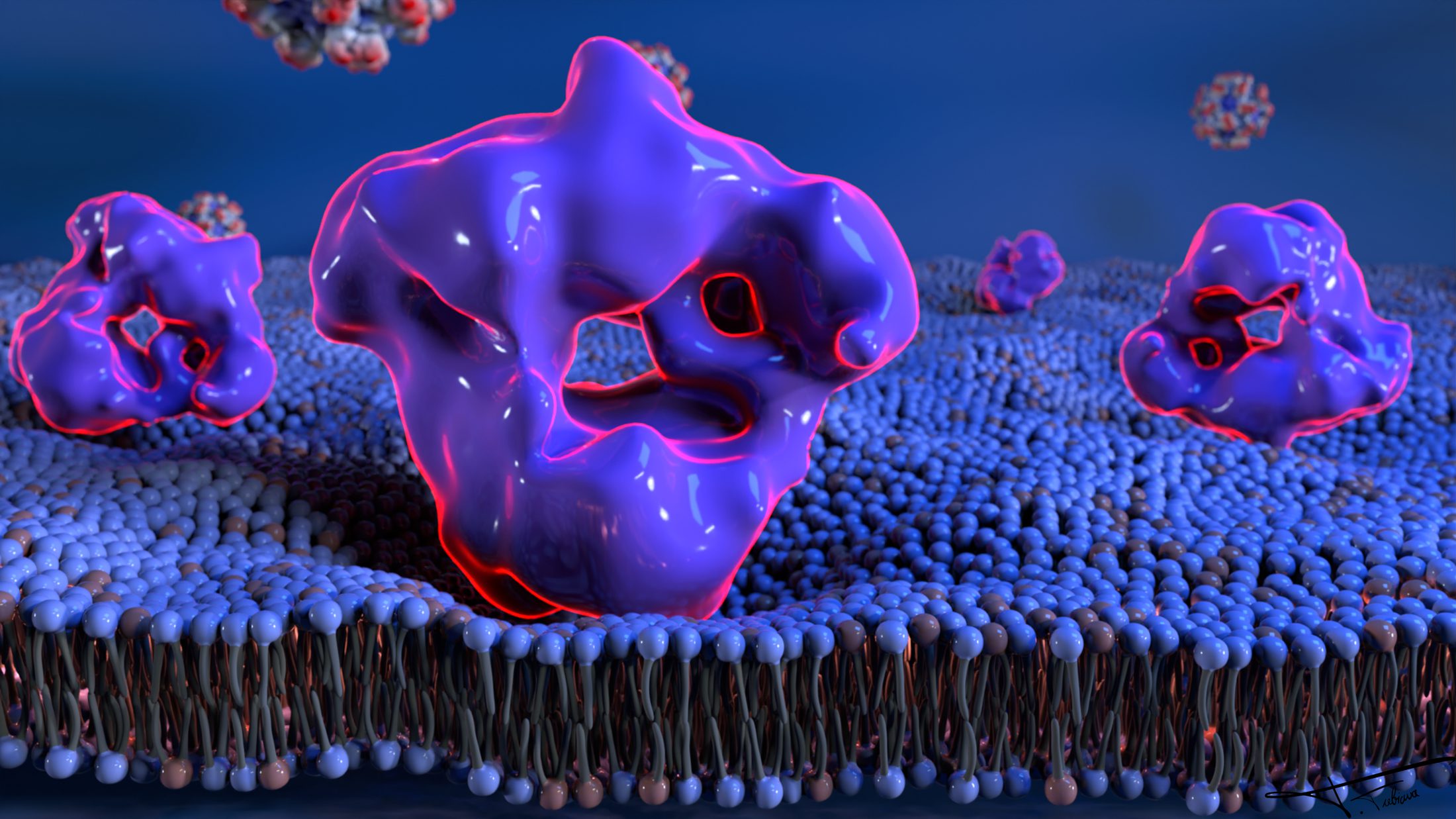
HEV ORF1 in a membrane
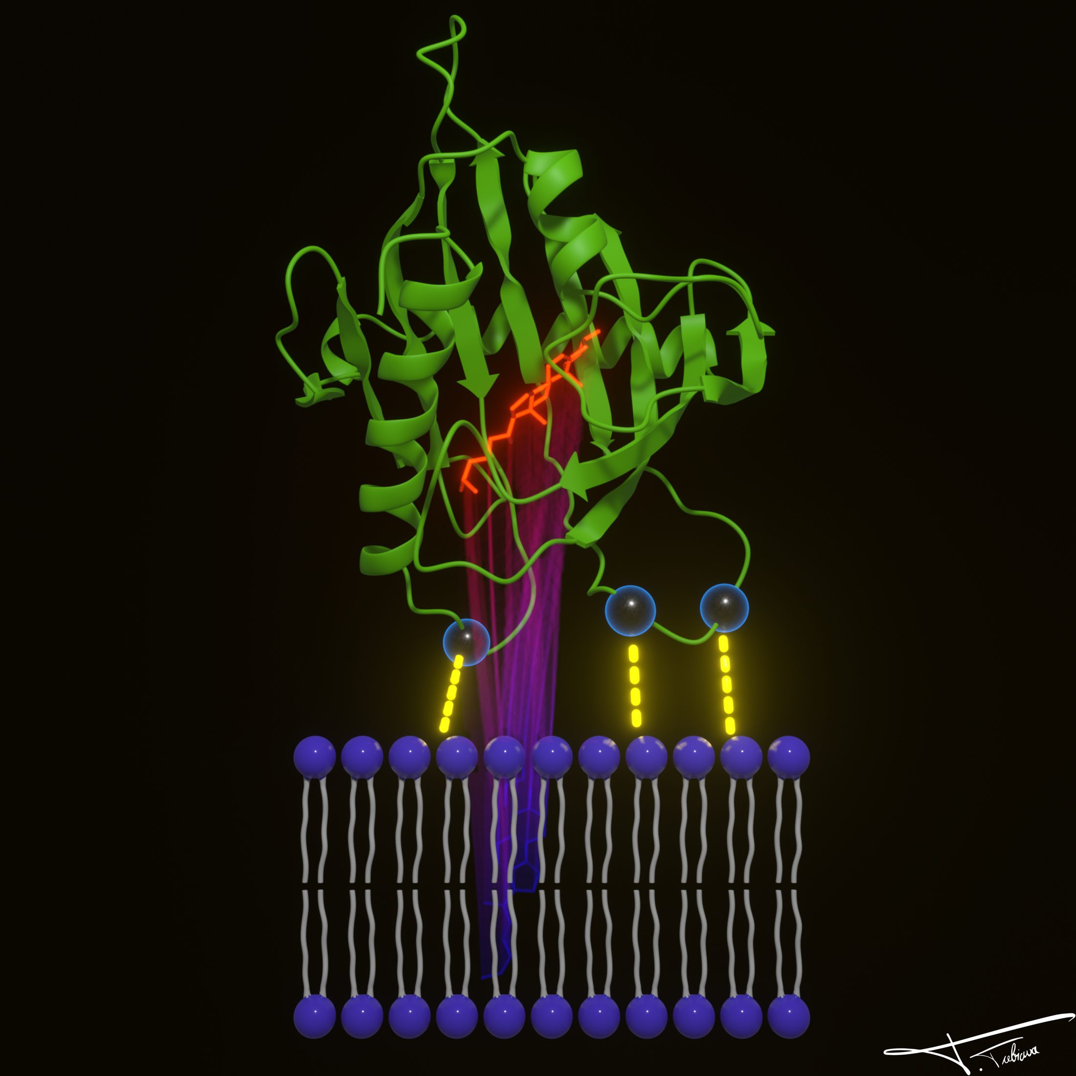
HEV ORF1 in a membraneThe replication polyprotein embedded in a lipid bilayer.
Molecular complexA multi-colored assembly, in motion.
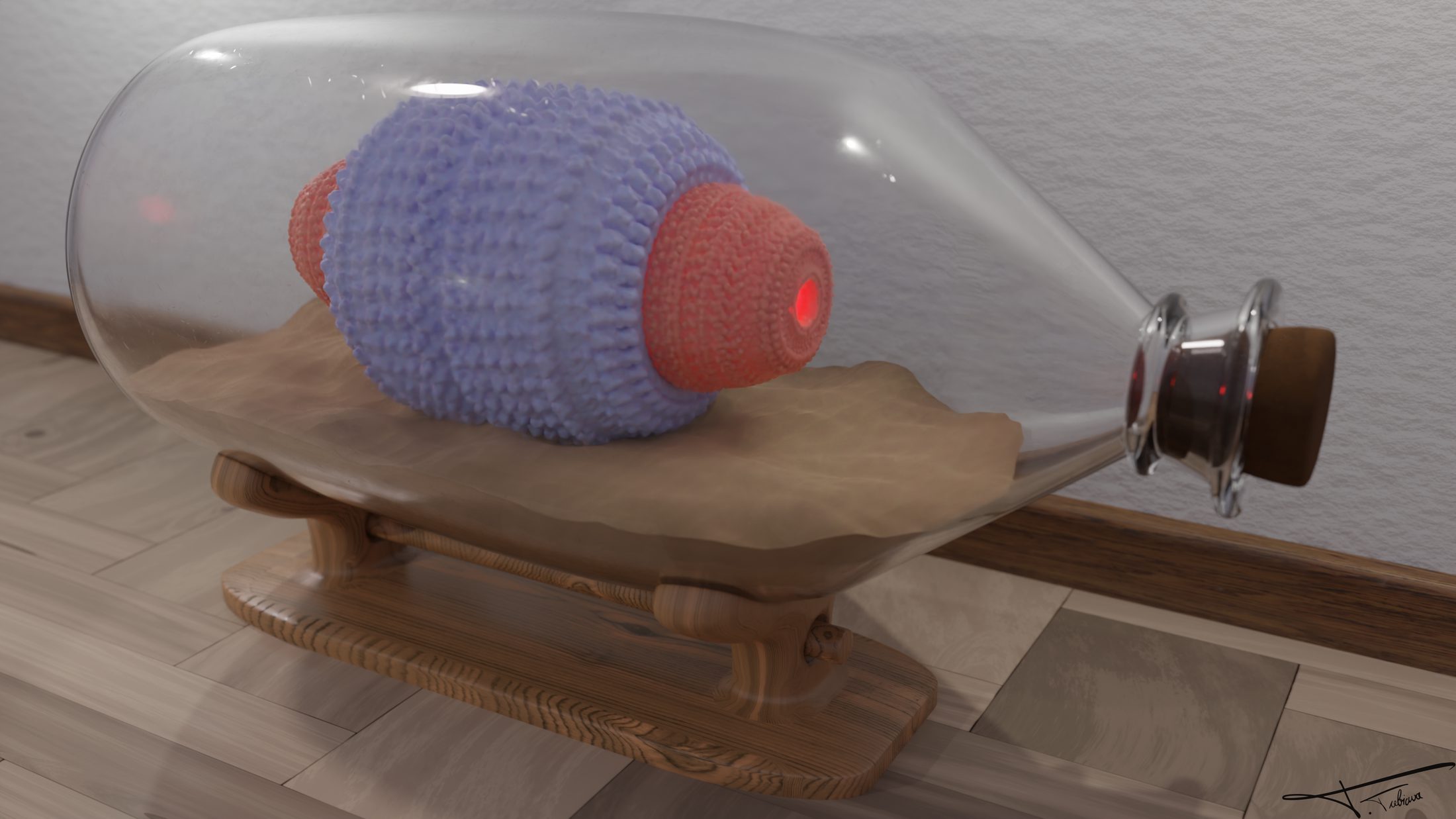
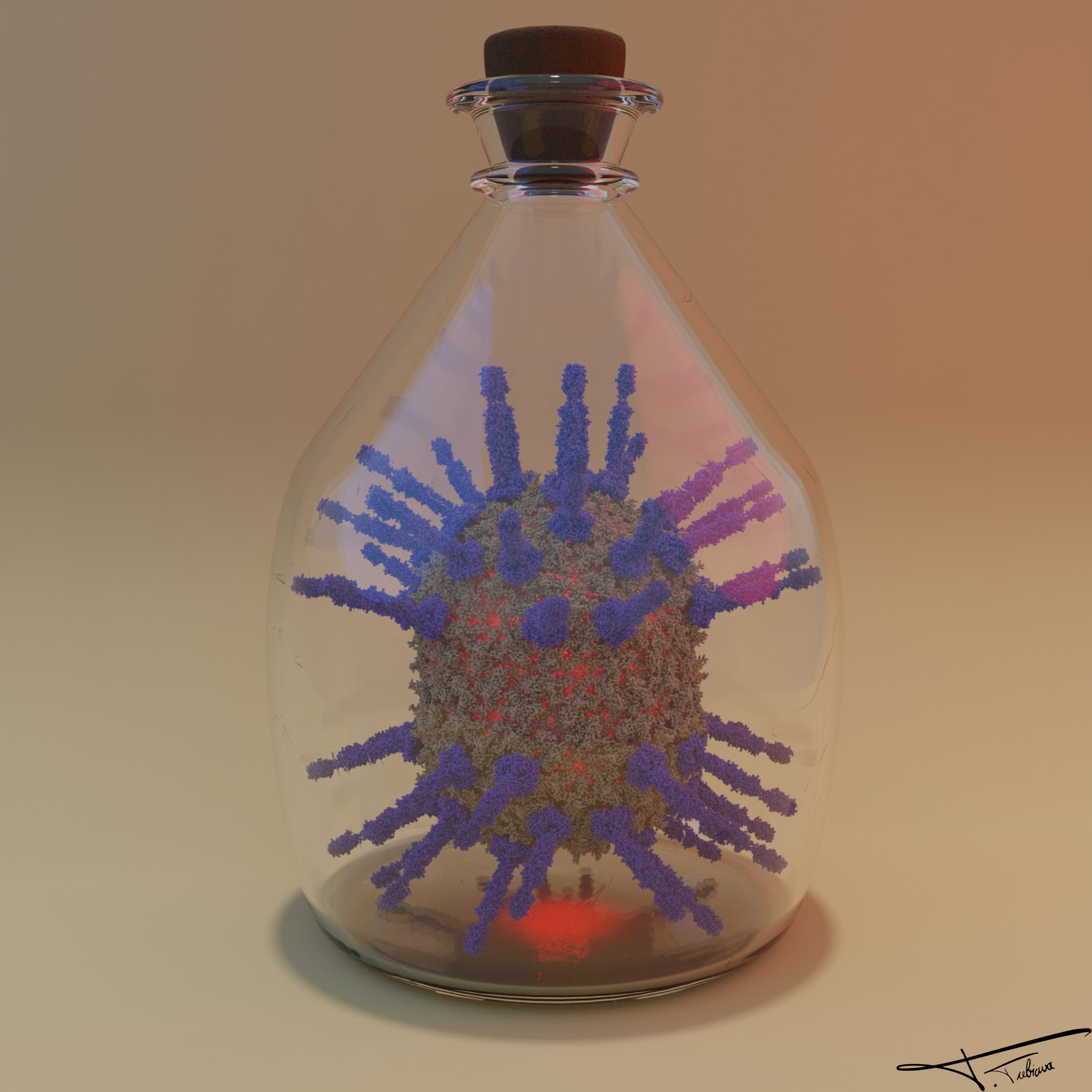
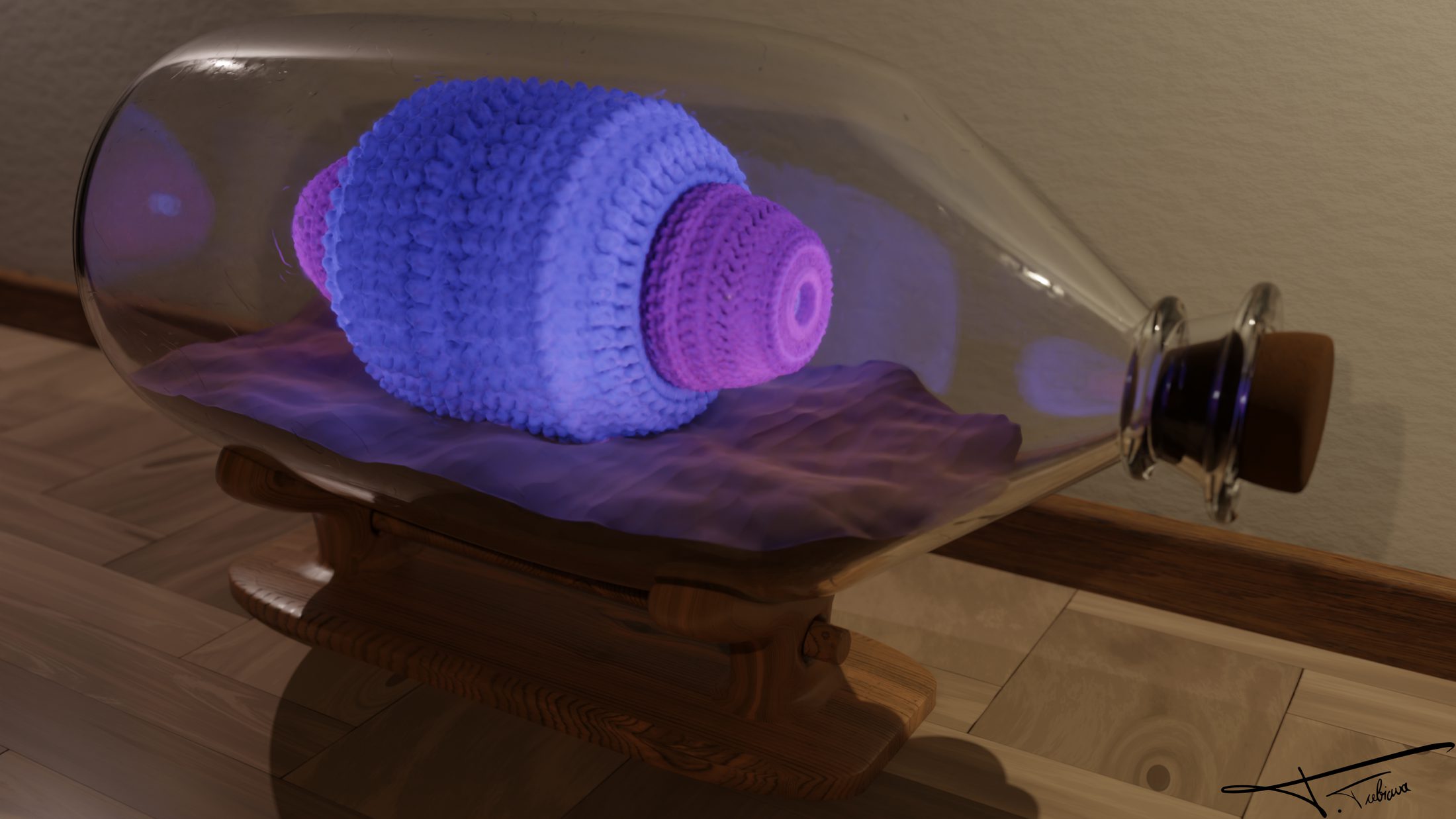
Virus in a bottle β€” PDB 6QZ0
Virus in a bottle β€” PDB 6QZ0A spiked particle, corked and displayed.
Union JackA flag, assembled atom by atom.
Membrane channel
Membrane channelA channel organizing itself inside a bilayer.
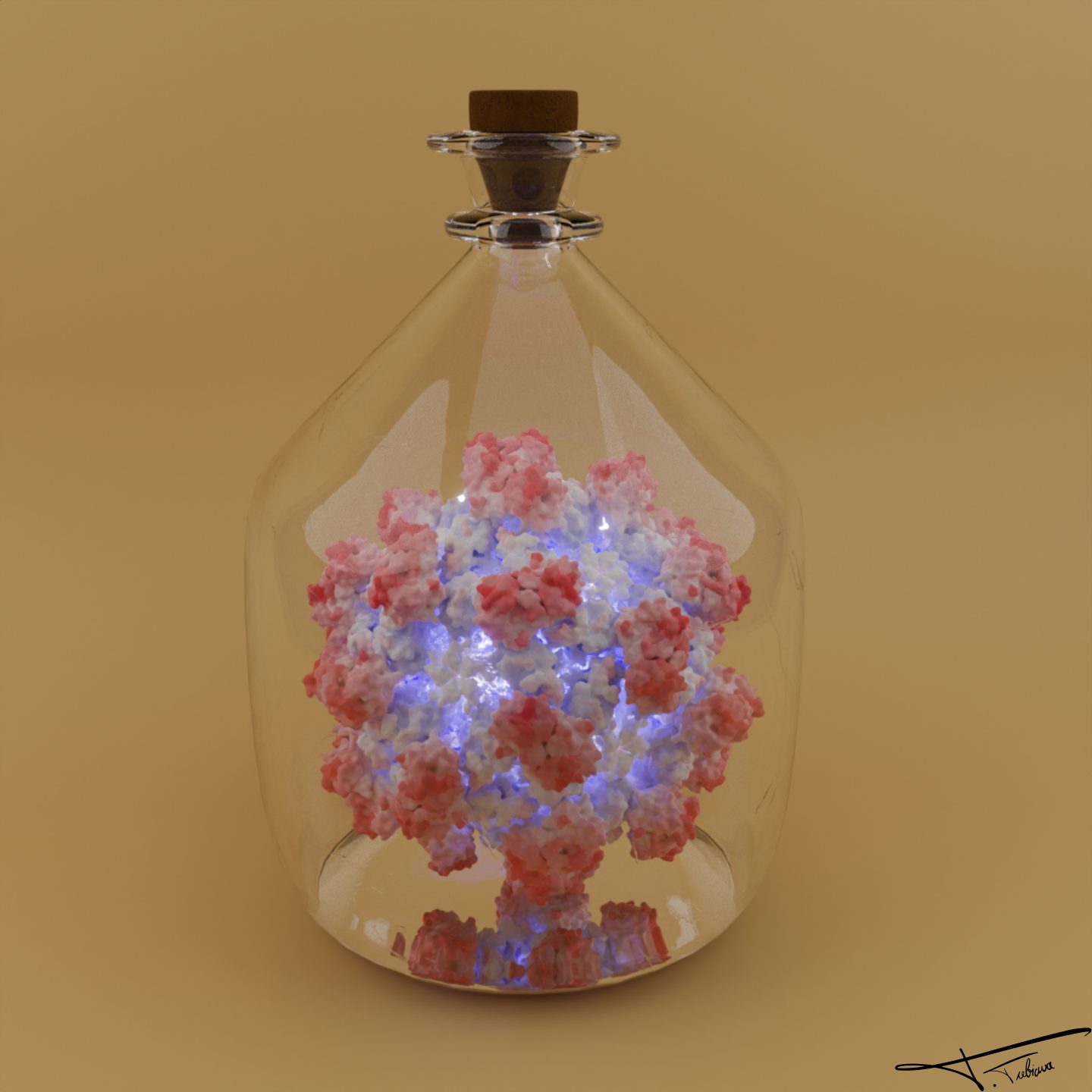
Hepatitis E in a bottle
Hepatitis E in a bottleA non-enveloped virus, kept on ice.
Membrane pore
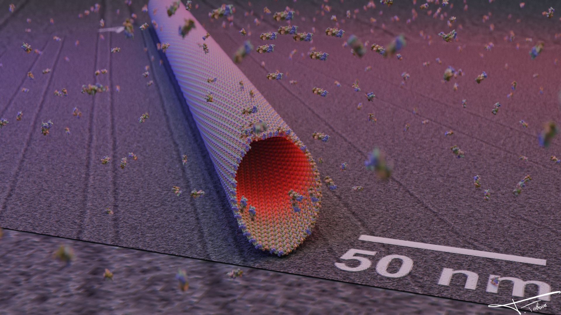
Membrane poreMolecular dynamics snapshot, 50 nm scale bar.
AlphaFold model β€” loopSeamless rotation of the same complex.
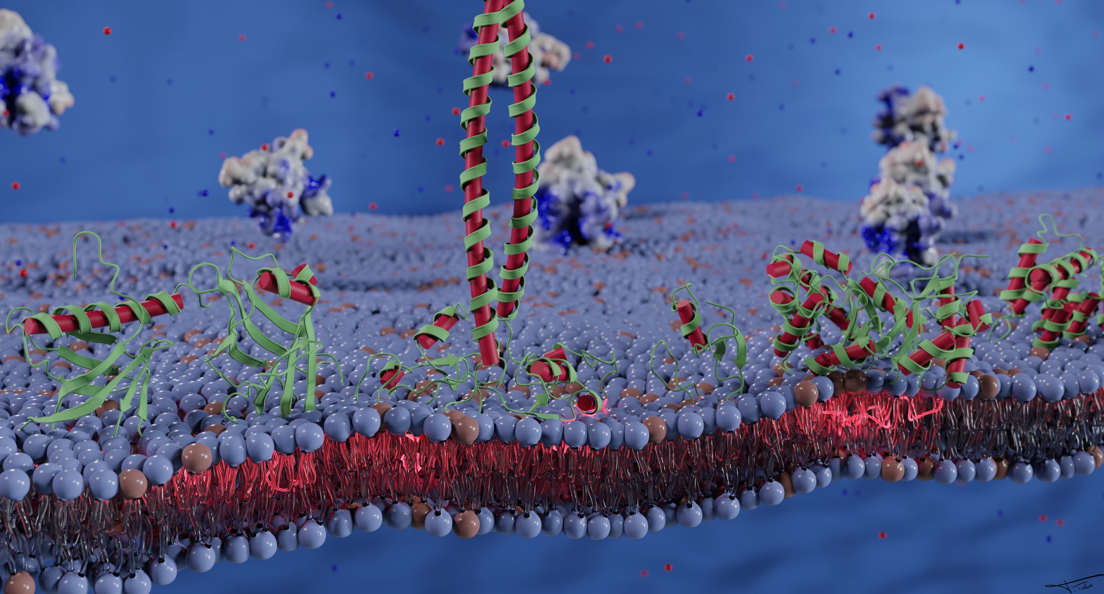
Cholesterol uptake
Cholesterol uptakeA membrane protein shuttling cholesterol.
A street at 50 nmAn urban scene, scaled to the molecular.
APOE3
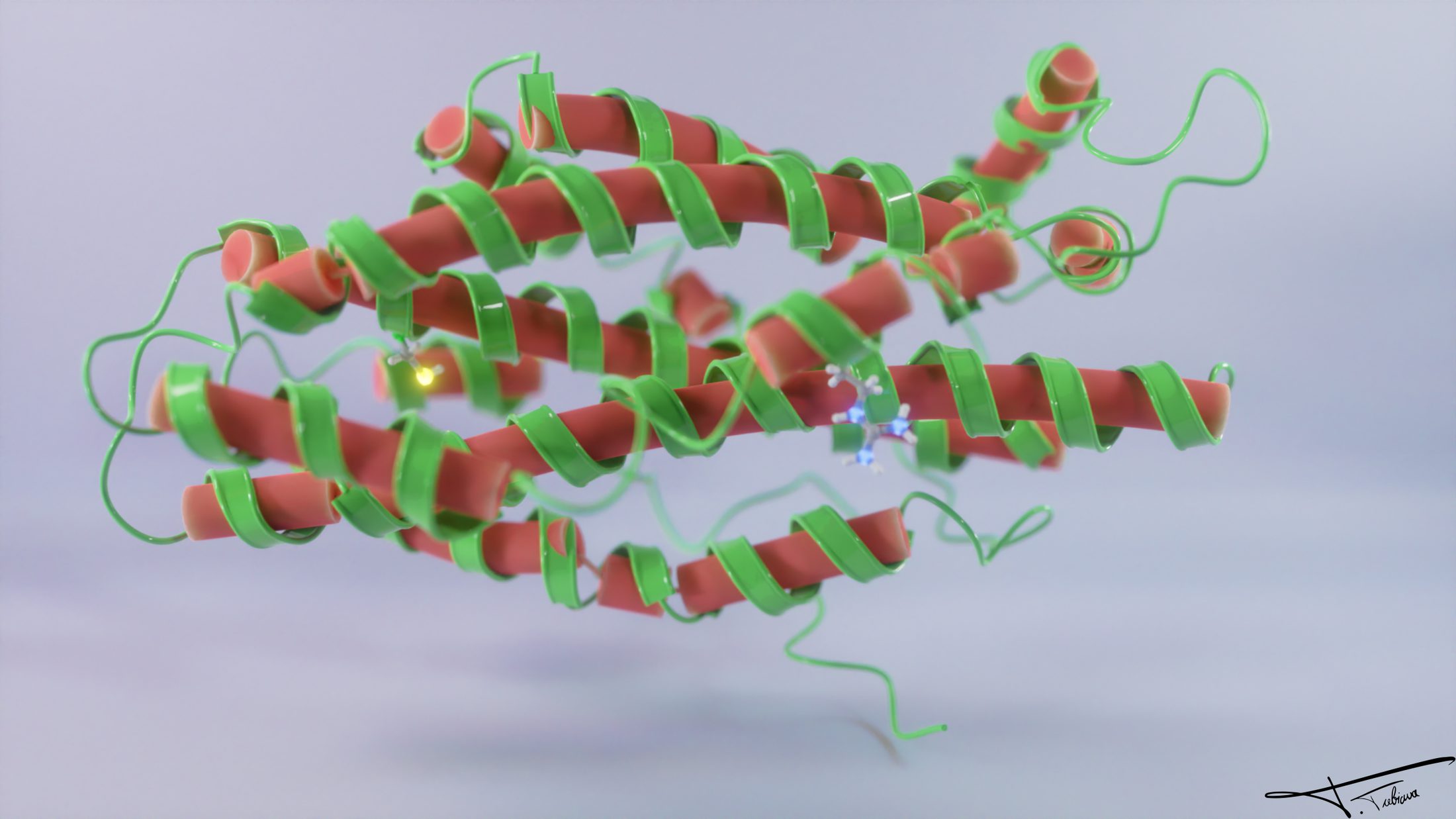
APOE3Secondary-structure coloring, with bound ligands.
Neon dodecamer β€” loopThe lighting sequence, on repeat.
Norovirus in a bottle β€” alternate
Norovirus in a bottle β€” alternateAnother take on the PDB 4V60 scene.
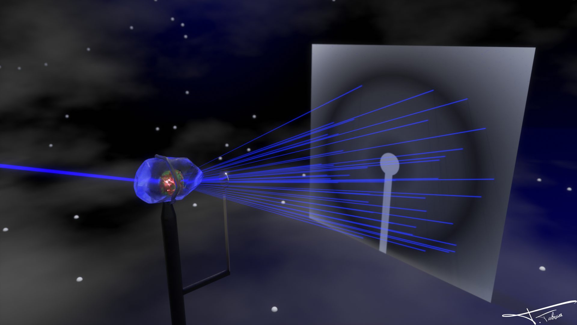
X-ray beam
X-ray beamA stylized diffraction experiment.
Protein complex β€” PDB 2NQ2
Protein complex β€” PDB 2NQ2A multi-subunit assembly on a neutral backdrop.
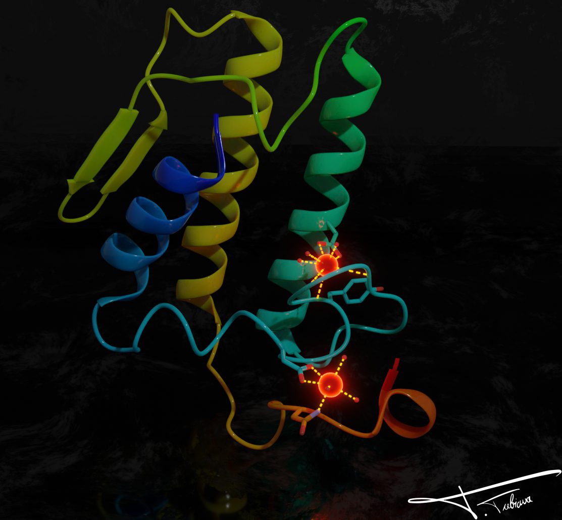
Transmembrane protein β€” PDB 1POA
Transmembrane protein β€” PDB 1POARibbon diagram with a bound ligand.
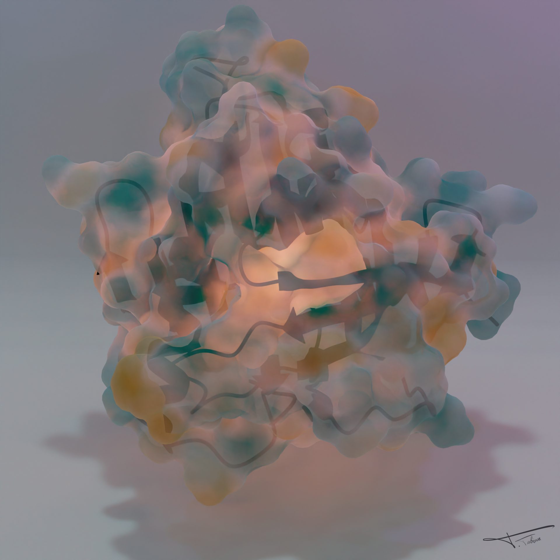
STARD4
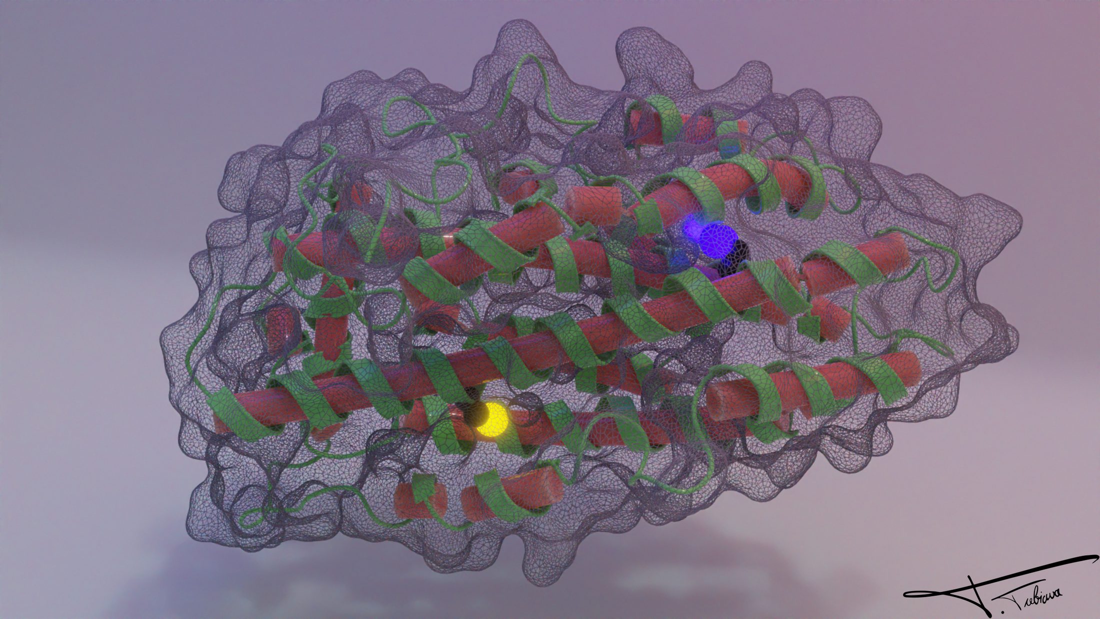
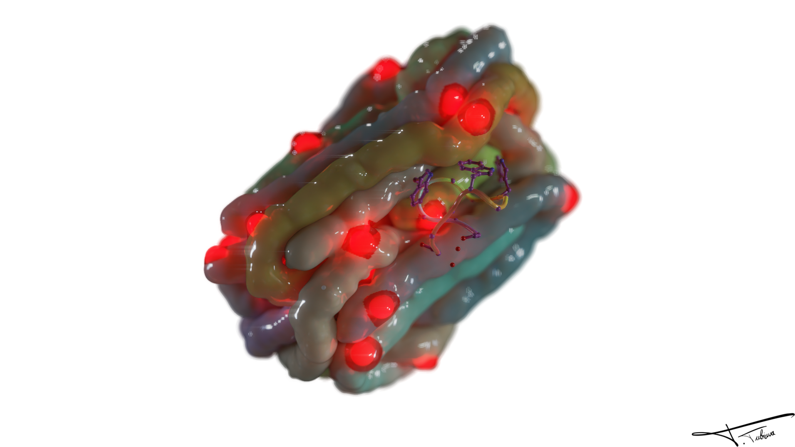
STARD4A translucent surface study.
APOE1
APOE1The apolipoprotein, with its density.
AlphaFold model β€” reversedPlayed in reverse, with an emphasis ring.
PDB 0233, Goodsell style
PDB 0233, Goodsell styleRendered in the iconic illustration style.
Viral capsid β€” variant
Viral capsid β€” variantAlternate coloring of the portal view.
Membrane, 512 GPU cycles
Membrane, 512 GPU cyclesA large bilayer, after a long render.
Neon dodecamer β€” lights offThe sequence, played backward.
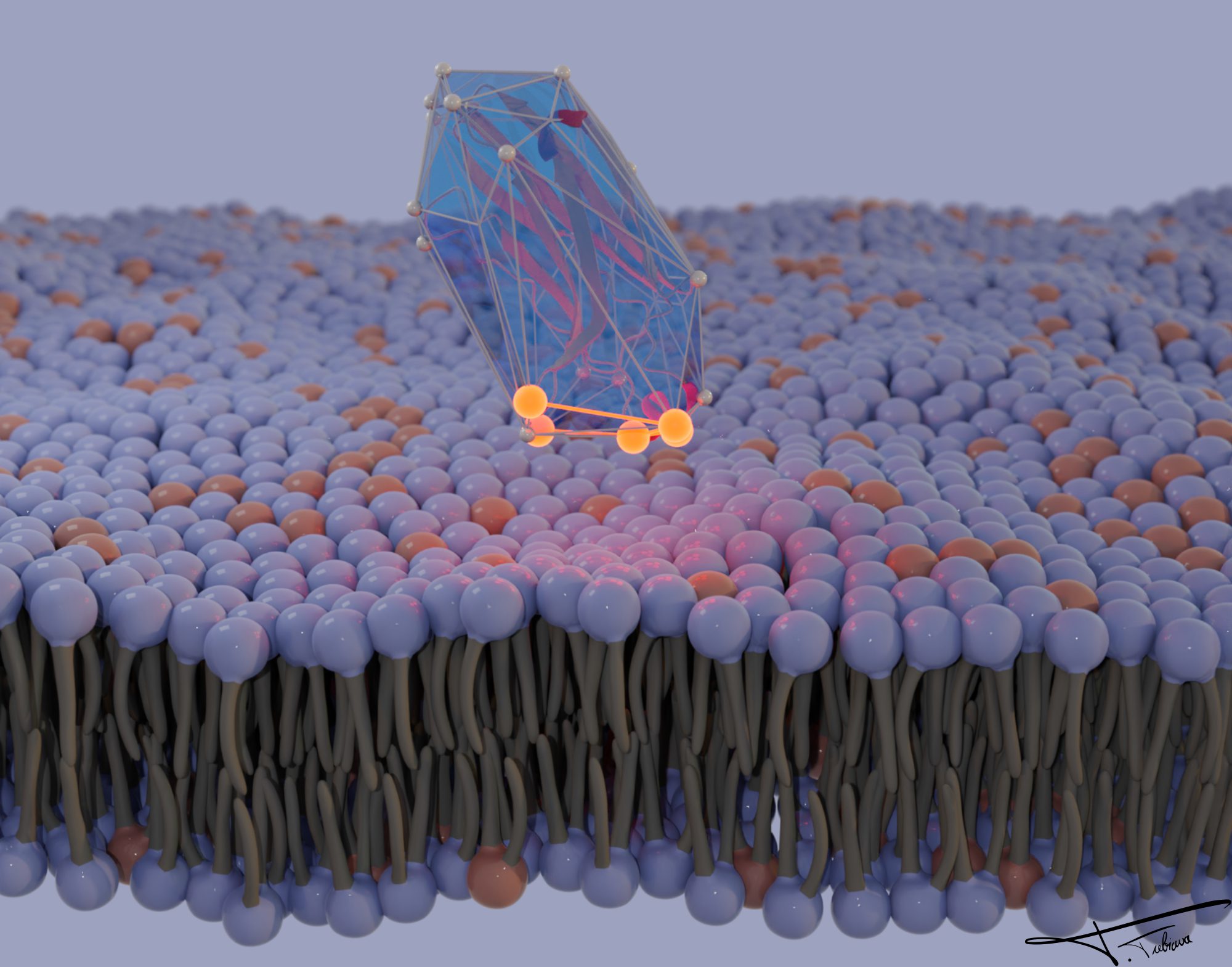
Convex hull
Convex hullA membrane region wrapped in its hull, with a crystal-like defect.
Material test 01
Material test 01A plastic shading study.
Material test 02
Material test 02Same idea, different light.
Simulation render
Simulation renderA frame from a longer study.
Electron density
Electron densityA density map brought to light.